English
  Español


Ask most women to point out the clitoris on a diagram and the finger lands in the right place immediately. Ask what shape the rest of it takes, and the answers thin out fast. That gap is not a lapse of attention. It is a lapse in the material: for most of the twentieth century, the diagrams handed to students showed a small labelled dot and nothing underneath it.
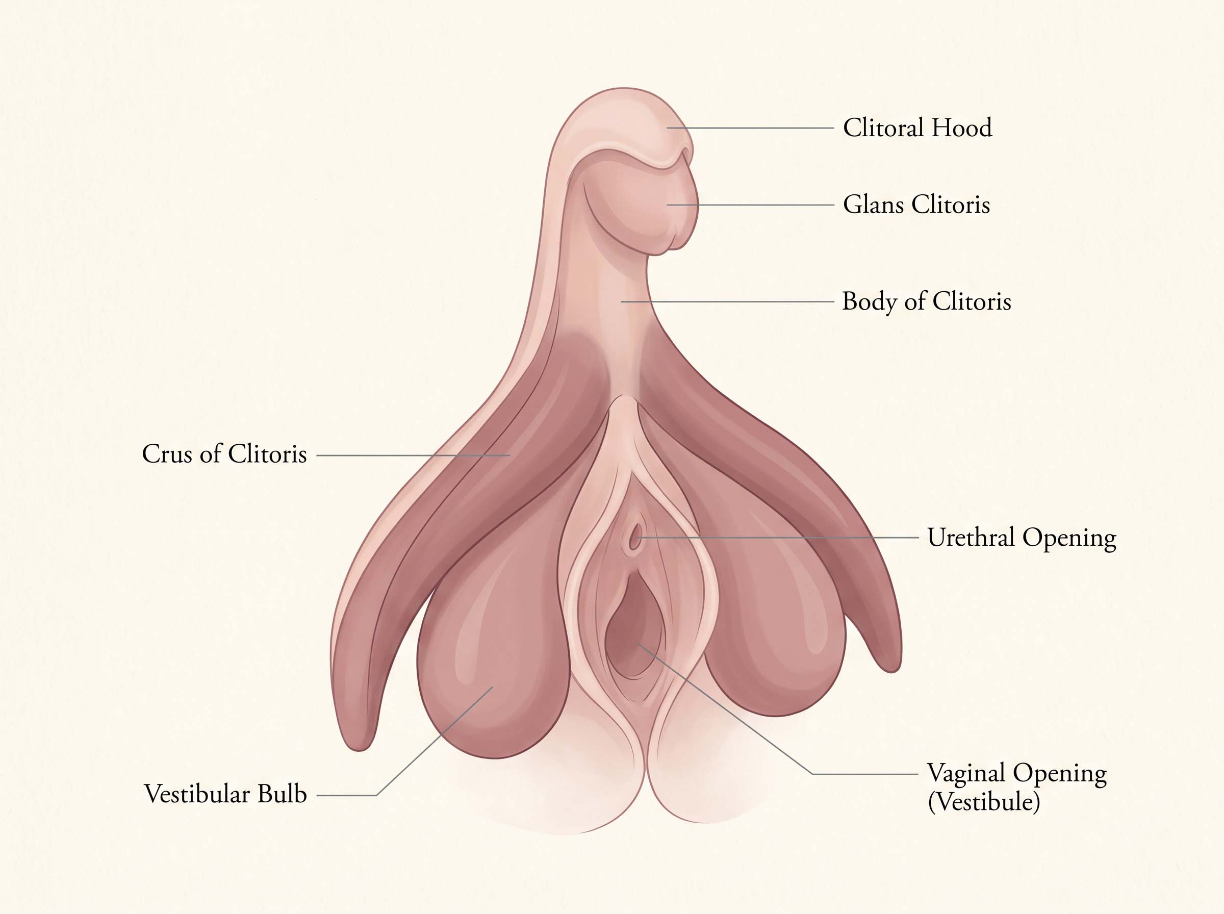
What sits underneath is larger and stranger than the label suggests. There is a body, a pair of arms that curve back into the pelvis, and two lobes of erectile tissue flanking the vaginal opening. Set against a full map of the vulva and its structures, the visible part reads as the tip of something roughly the shape of a wishbone — and the whole assembly is one organ, not a button with accessories.
The part everyone sees, and the part almost nobody draws
The visible portion is the glans, usually a few millimetres across, sheltered under a fold of skin called the hood. Behind it, running upward beneath the skin, is the body of the clitoris. The body then divides into two crura — the arms — which angle down and back along the pubic arch, each one several centimetres long in an adult woman. Alongside them sit the vestibular bulbs, two elongated cushions of erectile tissue that lie under the labia and along either side of the vaginal entrance.
All of it is built from the same class of tissue: spongy, richly supplied with blood vessels, and capable of swelling. Arousal fills that tissue, which is why the structure changes shape and firmness rather than staying fixed. Anatomists describe it as multiplanar — it does not lie flat in one drawing plane, which is one practical reason the older textbook illustrations reduced it to a dot. A single cross-section could not hold it.
One difference from its male counterpart is worth stating plainly, because it explains a great deal. The penis carries the urethra through its length; the clitoris does not. The urethral opening in women sits separately, below the glans. That single divergence in plumbing means the clitoris has no role in urination and no role in reproduction. Among human organs, that makes it unusual: no other structure in the body has sensation as its only known job.
No other structure in the human body has sensation as its only known job.
One blueprint, two outcomes
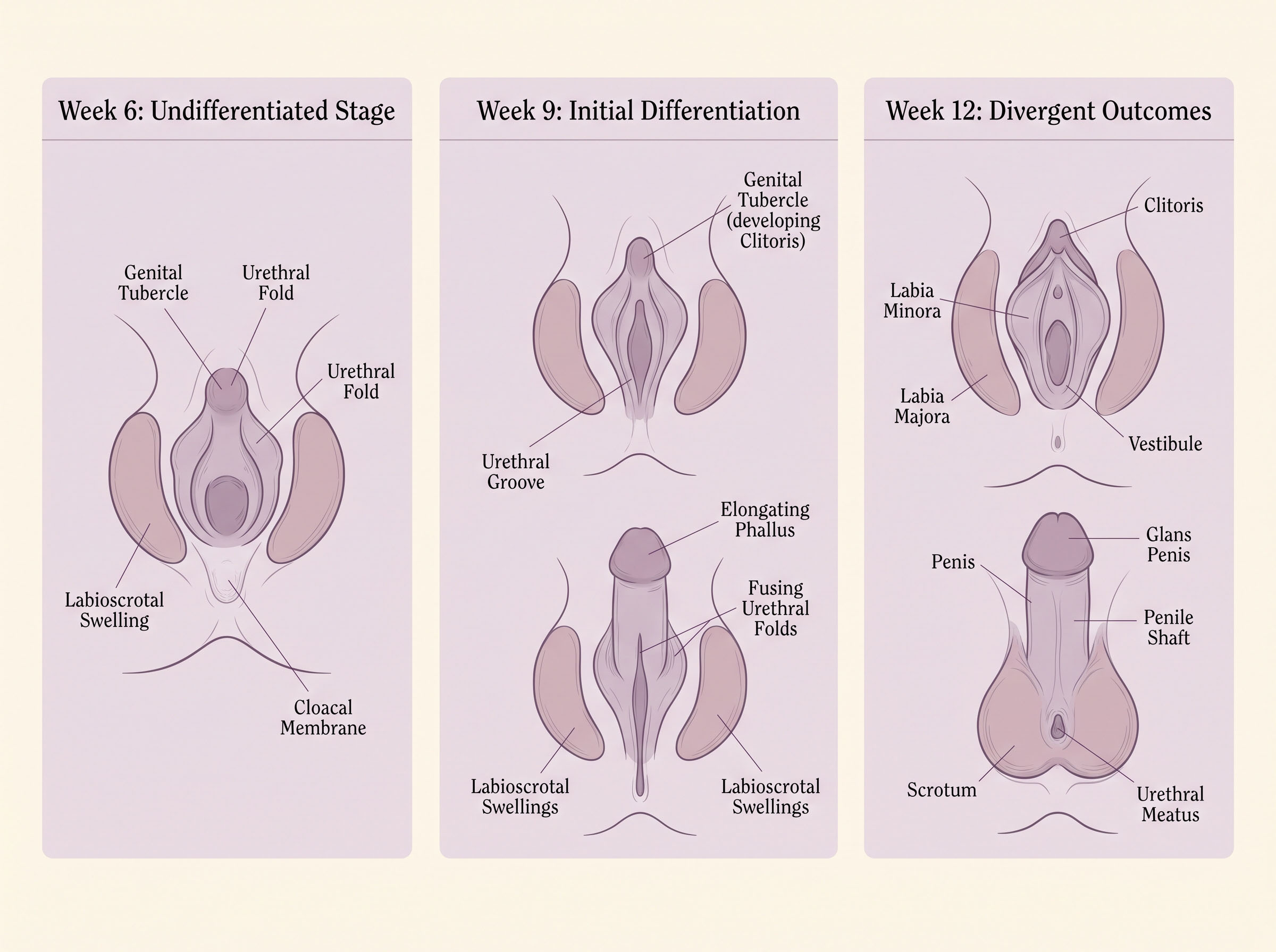
For the first weeks after conception, every human embryo is built to the same external plan. There is a small midline bump called the genital tubercle, a pair of folds on either side of it, and a pair of swellings outside those. Nothing about that arrangement is male or female yet. It is a single starting kit.
Around the seventh to ninth week, hormonal signalling decides how the kit is assembled. Where strong androgen signalling is present, the tubercle elongates, the folds fuse along the midline and enclose the urethra, and the outer swellings join to form the scrotum. Where that signalling is absent or low, the tubercle stays proportionally small and becomes the clitoral glans, the folds remain open and become the labia minora, and the outer swellings stay separate as the labia majora. The urethra keeps its own exit.
This is what anatomists mean by homology: not that one organ is a version of the other, but that both are made from the same embryonic material under different instructions. The erectile tissue is the same tissue. The hood and the foreskin are the same fold of skin. The ovary and the testis begin as the same gonad. Comparative biologists studying how androgens shape anatomy across mammals have used exactly this shared blueprint to work out how far hormone exposure can push a body plan in either direction.
| Embryonic origin | Develops into (female) | Develops into (male) |
|---|---|---|
| Genital tubercle | Clitoral glans and body | Glans and shaft of the penis |
| Urogenital folds | Labia minora (remain unfused) | Underside of the penile shaft (fused) |
| Labioscrotal swellings | Labia majora | Scrotum |
| Prepuce fold | Clitoral hood | Foreskin |
| Corpora cavernosa | Clitoral body and crura | Erectile columns of the penis |
| Urogenital sinus tissue | Vestibular bulbs | Corpus spongiosum |
| Indifferent gonad | Ovary | Testis |
The paper that redrew the map
By the 1990s the situation was odd. Surgical atlases described the male pelvis in fine detail; the female equivalent was thinner, and the clitoris in particular was often reduced to a single line. Helen O'Connell, who became Australia's first female urologist in 1993, noticed the discrepancy while studying and went looking for the missing material.
Her dissection work, published in 1998, examined how the urethra sits in relation to the surrounding erectile tissue and argued that the standard descriptions and the standard nomenclature were both inadequate. Seven years later she and her colleagues followed it with a full account in a 2005 review of clitoral anatomy in the Journal of Urology, drawing on dissection, magnetic resonance imaging of living women, histology and three-dimensional reconstruction. That review made two points at once: here is the structure, and here is the record showing that accurate descriptions had been available since the 1840s while textbooks in circulation in 2005 still omitted or misdescribed it.
Historians of anatomy have since treated the episode as a case study in how scientific knowledge can be lost without ever being disproved. It was not refuted. It was dropped.
Roughly nine tenths of the organ is internal. The glans that appears on a standard diagram represents only the exposed tip of a structure that extends several centimetres back into the pelvis on both sides.
Counting what had never been counted
For decades, popular articles repeated a figure: eight thousand nerve endings. The number was quoted so often it acquired the feel of a measurement. It was not one. Nobody had counted.
That changed with work carried out at Oregon Health & Science University and published in the Journal of Sexual Medicine in 2023. Researchers took samples of the dorsal nerve of the clitoris — the main nerve carrying sensory signals — stained the tissue, magnified it a thousand times, and counted the individual myelinated fibres under a microscope. The average came to about 5,140 fibres in a single dorsal nerve. Since the nerve is paired and symmetrical, the estimate for the pair worked out at roughly 10,281.
Two qualifications matter. The count covered myelinated fibres only, and the clitoris is served by additional smaller nerves that were not included, so the true figure is higher. And the sample was small — seven donors. What the study established is not a final number but a floor, and a method for anyone who wants to refine it.
5,140 — average myelinated fibres counted in one dorsal nerve of the clitoris.
10,281 — estimated total across the symmetrical pair, with a measured range of 9,852 to 11,086.
~18,000 — fibres in the median nerve, which serves a large portion of the hand. Fewer than twice the count, in a structure many times the size.
7 — tissue donors in the study, which is why researchers describe the result as a first estimate rather than a settled figure.
Source: Uloko, Isabey & Peters, Journal of Sexual Medicine, 2023.
Variation is the ordinary case
Measured dimensions vary widely between healthy women, and the research that documents this is thinner than it should be. One of the more frequently cited studies, published in BJOG in 2005 by Jillian Lloyd and colleagues at the Elizabeth Garrett Anderson Hospital in London, measured the external genitalia of fifty premenopausal women and reported exactly what the title suggested: "normality" turned out to unfold across a broad span rather than cluster around a single figure. The authors framed their findings as information women and clinicians could weigh when cosmetic genital surgery was under discussion.
A larger cohort study of 208 premenopausal women produced average measurements with substantial spread on either side. The figures below are means with standard deviations — the spread is the point, not the midpoint.
| Measurement | Mean (cm) | Standard deviation |
|---|---|---|
| Clitoral glans length | 0.87 | ± 0.21 |
| Clitoral glans width | 0.60 | ± 0.15 |
| Clitoral hood (prepuce) length | 2.05 | ± 0.48 |
| Distance from clitoris to urethral opening | 2.24 | ± 0.55 |
| Labia minora width (right / left) | 2.12 / 2.20 | ± 0.86 / ± 0.96 |
Position varies as much as size. In some women the glans sits well clear of the hood; in others the hood covers it almost entirely, and the distance from the glans to the urethral opening ranges over a couple of centimetres across a group. Research on the documented range of vulval shapes and sizes points the same way: the published averages describe a middle of a distribution that is wide at both ends, which is a very different thing from a template.
What is still argued about
Terminology remains contested. Some anatomists accept the vestibular bulbs as part of the clitoris; others hold that the bulbs are a separate structure and that expanding the definition blurs a useful distinction. Proposed composite terms — describing the clitoris, urethra and vagina as a single functional complex — have been criticised in the anatomical literature as claims running ahead of the evidence.
Imaging studies using ultrasound and MRI have examined how the internal portions shift and engorge during arousal, and some researchers have argued this explains reported differences in sensation between one form of stimulation and another. That work is suggestive rather than settled, drawn from small numbers of participants, and other specialists read the same images differently.
The thinner areas are easy to list. How much the unmyelinated fibres contribute to sensation is unquantified. How nerve density varies between women has not been mapped. Whether the structural differences documented in the measurement studies correspond to anything at all in experience is, on the current evidence, unanswered — the handful of studies that have looked found no clear relationship, but they were small.
The pattern across all of it is consistent. This is a structure that was drawn accurately in 1844, quietly dropped from the standard references, restored to the literature only at the end of the twentieth century, and given its first real nerve count in 2023. Most of what is known has been established within the lifetime of the women reading it, and the questions still open are basic ones. For an organ this old, that is a remarkably short research record — and a fairly good indication that the next twenty years of anatomy will have more to say.
Questions Readers Ask
Disclaimer: All content on this website—including articles, educational materials, and interactive calculators—is for informational, educational, and entertainment purposes only. The calculations, percentiles, and outputs generated by tools on this site are based on general statistical data and mathematical models; they do not constitute medical data, a clinical assessment, or a diagnosis.
Nothing contained on this website is a substitute for professional medical advice, diagnosis, or treatment. Always seek the guidance of a qualified healthcare professional or urologist with any questions you have regarding physical development, anatomy, or health conditions. Reliance on any information or tools provided by this website is solely at your own risk.