English
  Español


For most of the twentieth century, the largest structure in the human body devoted entirely to sensation went unmapped, unlabelled, and in several editions of the standard anatomy text, unmentioned altogether.
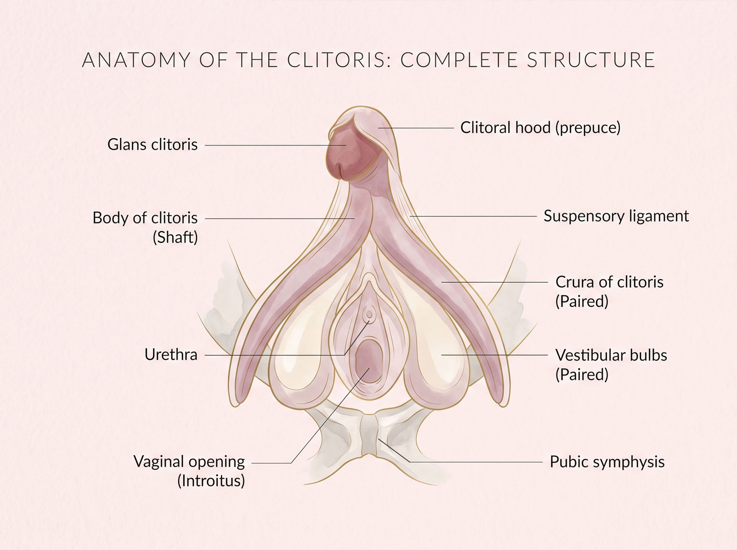
The full anatomical account did not appear in a major journal until 2005. Not the discovery of the organ — that had happened, and been forgotten, more than once. What arrived in 2005 was the complete architecture: the branching, mostly internal structure that anatomists had drawn in the nineteenth century and then let slip out of the textbooks for the better part of a hundred years. Anyone taught that the clitoris is a small button at the top of the vulva was taught roughly a quarter of the story. The rest sits beneath the surface, wrapping around the urethra and vaginal canal in a form that looks, in three dimensions, nothing like the diagram most women were shown in school. A fuller map of the vulva and its surrounding structures makes the scale of the omission clear.
A Structure Larger Than Its Reputation
Anatomists describe the organ as having several continuous parts. The glans is the visible portion, sheltered under a fold of tissue called the hood. Behind it, the body angles backward and divides into two arms — the crura — that run along the pubic bones. Alongside these sit the vestibular bulbs, paired masses of erectile tissue that lie on either side of the vaginal opening. Measured end to end in dissection studies, the whole complex commonly extends several inches, though published figures vary because anatomists have not always agreed on where one structure ends and the next begins.
What unites these parts is tissue type. Most of the organ is erectile, meaning it is built to fill with blood, and it is richly supplied with nerves. The 2023 histological work counting fibres in the dorsal nerve reported an average of roughly ten thousand myelinated fibres reaching the glans — considerably more than the figure of eight thousand that circulated in popular writing for decades, and which appears to have originated in a mid-century estimate derived from cattle rather than from human tissue.
The clitoris and the penis develop from the same embryonic tissue. Until roughly the ninth week of gestation, every human embryo carries an identical genital tubercle. Hormonal signalling then directs that shared starting point down one of two paths — which is why anatomists describe glans, shaft, erectile bodies and hood in both, and why the older idea that female anatomy was a lesser copy of male anatomy had the developmental sequence backwards.
The Long Century of Forgetting
The history is stranger than the anatomy. In 1559 the Italian anatomist Realdo Colombo announced the structure in print and claimed it as his own discovery; his contemporary Gabriele Falloppio promptly disputed the credit, and physicians in the Arabic medical tradition had described it centuries earlier still. In 1844 the German anatomist Georg Ludwig Kobelt produced illustrations of the full erectile complex — crura, bulbs and all — that hold up reasonably well against modern imaging.
Then the drawings thinned out. Across the late nineteenth and much of the twentieth century, editions of the leading anatomical reference reduced the organ to a labelled dot or dropped the label entirely, a pattern documented in the publishing history of Gray's Anatomy. Medical students trained in the 1970s and 1980s could complete a degree having read detailed chapters on male erectile function and nothing equivalent about the female counterpart.
| Year | Development | What Changed |
|---|---|---|
| 1559 | Colombo publishes a description and claims discovery | A dispute over credit begins that outlasts both men |
| 1844 | Kobelt illustrates the full erectile complex | The internal structures are drawn accurately — then set aside |
| 1905 | Freud proposes two categories of female orgasm | A theoretical claim reshapes clinical thinking for fifty years |
| 1966 | Masters and Johnson publish laboratory observations | Physiological recording replaces theory as the primary evidence |
| 1998 | O'Connell's dissection study on urethral relationships | Surgical relevance forces the question back into urology |
| 2005 | Full anatomical account published in the Journal of Urology | MRI and dissection findings are consolidated into one reference |
| 2009 | Sonographic imaging in the living state | Movement and engorgement observed rather than inferred |
| 2023 | Nerve fibres counted directly for the first time | A widely repeated popular figure is revised upward |
Sigmund Freud's 1905 proposal deserves its own line in that history. He argued that mature women transferred sensation away from the external organ, and that continued reliance on it signalled arrested development. No anatomical evidence supported the claim. It nonetheless shaped clinical practice for decades, and generations of women were told that ordinary physiology was a psychological failing. William Masters and Virginia Johnson's laboratory work in the 1960s recorded what actually happened in the body during arousal and found no basis for the division. The correction took another forty years to reach the textbooks.
"The organ was drawn correctly in 1844 and then quietly edited out of the books. What happened after that was not discovery. It was recovery."
What the Anatomy Suggests About Function
Biologists have argued about the evolutionary account for a long while, and the argument is not settled. One position holds that the structure is a by-product of shared embryonic development — present because the developmental programme that builds male erectile tissue runs, in modified form, in every embryo. A competing line of thinking treats it as functional in its own right, pointing to the density of nerve supply as evidence that selection acted on the structure directly rather than tolerating it as a leftover. Comparative work across other mammals has been used to support both readings, which is a fair sign that the evidence does not yet decide the matter. What can be stated without controversy is that the organ has no role in reproduction, urination, or any other bodily process. Among human anatomical structures, that singularity of purpose is unusual.
Two Islands, Two Silences
Mid-century anthropology produced two field studies that are still taught together. On Mangaia in the Cook Islands, ethnographers recorded a society in which the physical education of young men explicitly included responsibility for a wife's pleasure, discussed openly and instructed by elders.
In a small Irish community given the pseudonym Inis Beag, the same era of fieldwork described near-total silence on the subject within marriage, with the topic absent from conversation between mothers and daughters.
Both accounts have been criticised on methodological grounds since. What survives the criticism is the wider pattern anthropologists kept finding: reported experience tracks closely with whether a culture gives women the words for it.
Cross-cultural records reinforce how much the surrounding conversation matters. Physicians writing in the medieval Arabic and Persian traditions catalogued the structure without embarrassment as part of general medicine. Victorian English medical writing moved in the opposite direction — the physician William Acton famously asserted that respectable married women were largely untroubled by such feeling. The claim did not survive contact with data. When the American researcher Clelia Mosher quietly surveyed married women between the 1890s and 1920s, most of her respondents described desire, pleasure and a settled sense that these belonged inside marriage. Her findings sat unpublished in a Stanford archive until the 1970s, which says something about which version of Victorian womanhood was permitted into print.
Blood Flow, Engorgement, and the Question of Night-Time Cycles
Because most of the organ is erectile tissue, arousal produces measurable change: arterial supply increases, the tissue fills, and volume rises. Sonographic imaging in the living state has recorded this directly. The visible glans typically changes less dramatically than the internal portions, which is part of why the process was underestimated for so long — the observable part is the smallest part. Broader research into the role of blood flow in sexual and reproductive health covers the same vascular mechanics across other tissues.
This raises a question that comes up often and is rarely answered plainly: does anything comparable to a morning erection occur in women? Sleep-laboratory research from the 1970s and 1980s, conducted on small samples, recorded cyclical increases in genital blood flow during REM sleep in women — occurring on a rhythm broadly similar to the well-documented cycles measured in men, and likewise unconnected to dreaming content. The findings have been replicated rarely, the sample sizes were modest, and the phenomenon is far less studied than its male equivalent. It is not visible in the way the male version is, which is one reason it drew less attention and less funding. As a research area it remains thin, and honest summaries say so.

Pleasure, Marriage, and What the Research Actually Measures
A substantial body of survey research has examined how sexual satisfaction relates to relationship quality. Findings across large samples consistently show the two travelling together — couples who report one tend to report the other. What the research cannot settle is direction. A contented marriage may produce satisfying intimacy; satisfying intimacy may steady a marriage; a third factor, such as the quality of everyday communication, may drive both. Studies that follow couples over time suggest the influence runs in both directions, which is the least tidy answer and probably the most accurate one.
One pattern from large surveys is worth stating plainly, because it cuts against the assumption that this is primarily a matter of technique or anatomy. Reported frequency of orgasm among women in heterosexual relationships varies enormously by context, and the widest surveys find the difference between men and women narrows considerably within long-term committed partnerships compared with casual encounters. Familiarity, the freedom to say what is wanted, and time appear in the data as strongly as anything physiological. Researchers who study what happens in the body and brain during orgasm have described a cascade involving muscle activity, hormone release and altered blood pressure, though the mapping of subjective experience onto those measurements remains incomplete.
| ~10,000 | Average myelinated nerve fibres reaching the glans, per the 2023 histological count |
| 1844 | Year the full internal structure was accurately illustrated |
| 161 | Years between that illustration and the consolidated 2005 anatomical account |
| ~1/4 | Rough proportion of the organ that is externally visible |
What Remains Unsettled
Plenty. The evolutionary question has no consensus. Night-time engorgement cycles rest on a handful of small studies. Whether the vestibular bulbs should be classified as part of the organ or as neighbouring structures is still argued among anatomists, and the terminology in published papers reflects that disagreement. The relationship between the anterior vaginal wall, the urethra and the internal portions has been described differently by different research groups, with one line of work concluding that the much-discussed "G-spot" is not identifiable as a discrete structure on dissection. Funding patterns explain some of the gap. Research into male erectile function attracted commercial investment on a scale that female physiology never matched, and the resulting literature is correspondingly deeper. The 2005 review and the 2023 nerve count were both, in their authors' descriptions, responses to an absence — work undertaken because the basic reference material a surgeon or a clinician might reach for simply did not exist.
For a structure this well supplied with nerves, sitting in plain view of every anatomist for five hundred years, that absence remains the most remarkable fact in the whole record. The anatomy was never the difficult part.
Primary sources for this article include O'Connell, Sanjeevan & Hutson, "Anatomy of the Clitoris," Journal of Urology (2005), and Uloko, Isabey & Peters, the 2023 histomorphometric nerve-fibre study in The Journal of Sexual Medicine.
Common Questions About Clitoral Anatomy
Disclaimer: All content on this website—including articles, educational materials, and interactive calculators—is for informational, educational, and entertainment purposes only. The calculations, percentiles, and outputs generated by tools on this site are based on general statistical data and mathematical models; they do not constitute medical data, a clinical assessment, or a diagnosis.
Nothing contained on this website is a substitute for professional medical advice, diagnosis, or treatment. Always seek the guidance of a qualified healthcare professional or urologist with any questions you have regarding physical development, anatomy, or health conditions. Reliance on any information or tools provided by this website is solely at your own risk.